

Starting from the 1950s to the current times, a number of tridimensional wax reproductions of the vertebrate lymphatic vessels have been realized as a result of pioneering studies by the anatomists, Gaetano Ottaviani and Giacomo Azzali, working at the University of Parma. In particular, ultrastructural research by Giacomo Azzali has provided the basis for the development of an original ceroplastic technique.
Briefly, using photographic images of a single lymphatic vessel isolated from serially cut ultrathin sections and studied with transmission electron microscopy, it has been possible to prepare lithographic-like replicas of its section profiles. After deposition of a layer of wax onto the photographic image of the vessel section, each physical contour of the vessel profile can be isolated by surgical excision of the wax and the various contours piled one over the other to reconstruct the tridimensional wall of the lymphatic vessel under study.
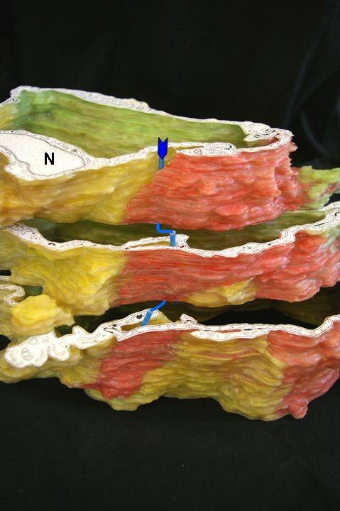
In this manner it has been possible to reproduce on a macroscopic scale the movement of immunocompetent (lymphocytes) and tumoral (metastases) cells from the interstitial to the intravascular compartment. Lymphocytes and metastases plastically deform the endothelial cell membrane and cytoplasm by "excavating" a penetrating channel called intraendothelial channel (arrow in figure), to give rise to the so-called lymphatic intravasation (1, 2). Intraendothelial channels spontaneously close after the passage of the moving cellular elements, without leaving any permanent interruption of the vessel wall.
These channels have astonishing similarities with anatomical structures predicted three centuries ago by the anatomist, Paolo Mascagni, referred to as "vascular pores" in his late publications (3).